


Stroke
Introduction and Facts
Stroke is a functional brain disorder, both focal and global acute, more than 24 hours, originating from disturbances in cerebral blood flow and not caused by transient cerebral blood circulation disorders, brain tumors, secondary strokes due to trauma or infection (WHO MONICA, 1986). 1996/1997 got 2,065 stroke patients from 28 hospitals in Indonesia (Misbach, 2000). The Indonesian Ministry of Health's survey on 987,205 subjects from 258,366 households in 33 provinces found that stroke was the leading cause of death at the age > 45 years (15.4% of all deaths). The average stroke prevalence was 0.8%, the highest was 1.66% in Nangroe Aceh Darussalam, and the lowest was 0.38% in Papua (RISKESDAS, 2007).
Pathophysiology
Infarction: Infarct stroke occurs due to a lack of blood flow to the brain. Normal blood flow to the brain is 58 mL/100 grams of brain tissue per minute; if it drops to 18 mL/100 grams of brain tissue per minute, the electrical activity of the neurons will stop even though the cell structure is still good, so the clinical symptoms are still reversible. If blood flow to the brain falls to <10 mL/100 grams of brain tissue per minute, a series of irreversible cell and membrane biochemical changes will occur, forming an infarcted area.
Intracerebral hemorrhage: Approximately 10% of strokes are caused by intracerebral bleeding. Hypertension, especially uncontrolled, is a significant cause. Other causes include ruptured aneurysms, arteriovenous malformations, cavernous angiomas, alcoholism, blood dyscrasias, anticoagulant therapy, and amyloid angiopathy.
Subarachnoid hemorrhage: Most cases are caused by rupture of an aneurysm in the branching of the great arteries.
Other causes are arteriovenous malformations or tumors.
Clinical Symptoms and Complications
Any stroke will cause acute neurological deficits (De Freitas et al., 2009), which include:
- Sensory hemideficit,
- Loss of consciousness,
- Central facial (VII) and hypoglossal (XII) nerve paralysis, impaired sublime functions such as language difficulties (aphasia) and intellectual function disorders (dementia),
- Blind half of the field of view (hemianopsia),
- Brainstem deficit.
Diagnosis
The diagnosis of stroke is made based on the history, clinical symptoms, and investigations. Laboratory examinations play a role in several things, including ruling out other neurological disorders, detect the cause of stroke, and find comorbid conditions.
Medication and Treatment
HYPERACUTE STADIUM
Actions at this stage are carried out in the Emergency Room and are cerebro-cardiopulmonary resuscitation measures to prevent brain tissue damage from spreading.
The patient is given oxygen at 2 L/min and crystalloid/colloid solutions; avoid giving dextrose fluids or saline in H2O. No CT scan, electrocardiography, chest X-ray, complete peripheral blood and platelet count, prothrombin time/INR, APTT, blood glucose, blood chemistry (including electrolytes) examinations were performed. If hypoxic, blood gas analysis is performed. Another action in the Emergency Room is to provide mental support to patients and explain to their families to stay calm.
Ischemic Stroke
General therapy: Place the patient's head in a 300 position, head and chest in one plane; change sleeping position every 2 hours; Gradual mobilization when hemodynamics is stable. Next, clear the airway and give oxygen 1-2 liters/minute until blood gas analysis results are obtained. If necessary, intubate. Fever is treated with compresses and antipyretics, then look for the cause; if the bladder is full, emptied (preferably by intermittent catheter).
Provide nutrition with 1500-2000 mL isotonic, crystalloid or colloid fluids and electrolytes as needed, avoiding fluids containing glucose or isotonic saline. Give food orally only if the swallowing function is good; If swallowing problems or loss of consciousness are found, a nasogastric tube is recommended. If increased intracranial pressure is found, intravenous bolus mannitol is given 0.25 to 1 g/kg body weight per 30 minutes. If rebound phenomenon is suspected or general condition worsens, 0.25 g/kg body weight per 30 minutes every 6 hours for 3-5 days is suspected. Osmolality monitoring should be performed (<320 mmol); alternatively, a hypertonic solution (3% NaCl) or furosemide can be given.
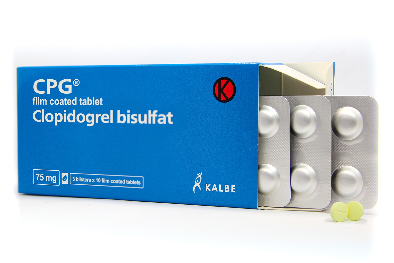
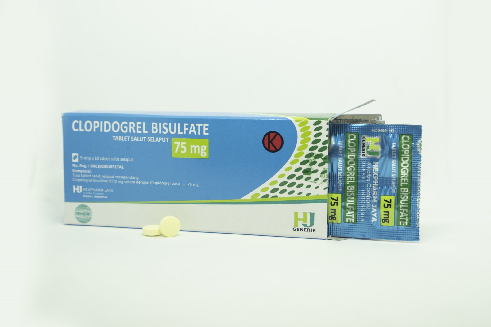
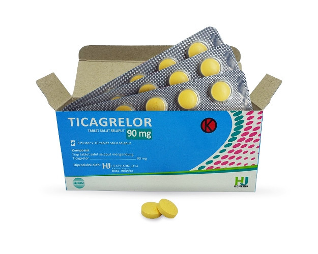
Specific therapy: Intended for reperfusion with the administration of antiplatelets such as aspirin and anticoagulants, or recommended with the thrombolytic rt-PA (recombinant tissue Plasminogen Activator). Neuroprotective agents can also be given, namely citicoline or piracetam (if aphasia is obtained).
Hemorrhagic Stroke
General therapy: Patients with hemorrhagic stroke should be admitted to the ICU if the hematoma volume is >30 mL, intraventricular hemorrhage with hydrocephalus, and the clinical condition worsens. Blood pressure should be reduced to premorbid blood pressure or 15-20% if systolic pressure >180 mmHg, diastolic >120 mmHg, MAP >130 mmHg, and hematoma volume increases.
If heart failure is present, blood pressure should be lowered immediately with labetalol iv 10 mg (2 min administration) to 20 mg (10 min administration) maximum 300 mg; enalapril i.v. 0.625-1.25 mg every 6 hours; captopril three times 6.25-25 mg orally. If there are signs of increased intracranial pressure, head position raised 30 degrees, head and chest position in one plane, administration of mannitol (see management of ischemic stroke), and hyperventilation (pCO2 20-35 mmHg).
General management is the same as for ischemic stroke, gastric ulcers treated with parenteral H2 antagonists, sucralfate, or proton pump inhibitors; Airway complications are prevented by physiotherapy and treated with broad-spectrum antibiotics.
Specific therapy: Neuroprotectors can be given except those that are vasodilators. Surgery considers the age and location of the bleeding, namely in patients whose condition is getting worse with cerebellar bleeding >3 cm in diameter, acute hydrocephalus due to intraventricular or cerebellar bleeding, VP-shunting, and lobar bleeding >60 mL with signs of acute increased intracranial pressure and the threat of herniation.
In subarachnoid hemorrhage, calcium antagonists (nimodipine) or surgery (ligation, embolization, extirpation, or gamma knife) may be used if the cause is an aneurysm or arteriovenous malformation (AVM).
SUBACUTE STADIUM
Medical action can be cognitive therapy, behavior therapy, swallowing, speech therapy, and bladder training (including physical therapy). Due to the long course of the disease, intensive exceptional post-stroke management is needed in hospitals to ensure patient independence, understanding, understanding, and implementing primary and secondary preventive programs.
Subacute phase therapy:
- Continuing therapy according to the previous acute condition,
- management of complications
- Restoration/rehabilitation (according to patient needs), namely physiotherapy, speech therapy, cognitive therapy, and occupational therapy,
- Secondary prevention
- Family Education and Discharge Planning
Reference:
Setyopranoto I. Stroke: Symptoms and management